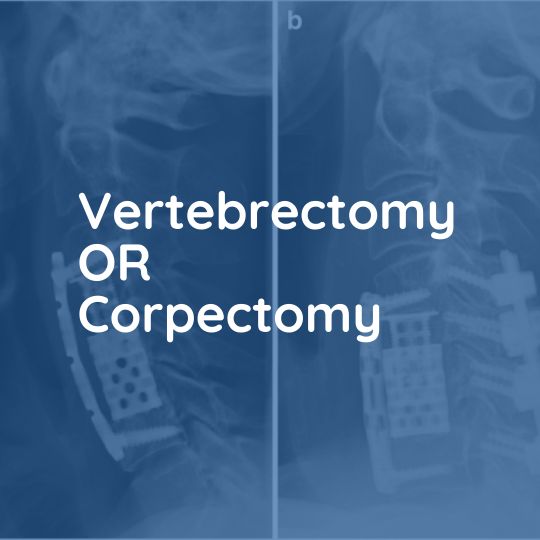
Vertebrectomy/Corpectomy
Vertebrectomy or corpectomy is a surgical procedure that involves the removal of a vertebral body in the spine. This procedure is usually performed to relieve pressure on the spinal cord or nerve roots due to a tumor, fracture, or other condition.
Causes
Symptoms
Treatment
Causes
The symptoms of spinal cord or nerve root compression can vary depending on the location and severity of the compression, but may include pain, numbness or tingling, weakness, difficulty with coordination, and problems with bowel or bladder function.
Symptoms
The symptoms of spinal cord or nerve root compression can vary depending on the location and severity of the compression, but may include pain, numbness or tingling, weakness, difficulty with coordination, and problems with bowel or bladder function.
Treatment
Vertebrectomy or corpectomy is usually performed as a last resort when other treatments such as medication, physical therapy, or injections have not provided relief. The procedure involves removing the affected vertebra and replacing it with a bone graft, artificial implant, or other material to stabilize the spine and relieve pressure on the spinal cord or nerve roots.
Who Needs It?
Vertebrectomy is performed either when there is abnormality affecting the bone of the vertebral body, such as a tumor deposit, or when there is pathology affecting the disc above and below one bone and the disc material is protruding behind the body of the intervening vertebra and is calcified e.g in Ossification of the posterior longitudinal ligament (OPLL).
The Operation
This is performed in exactly the same manner as an anterior cervical discectomy and fusion, through a small incision in the front of the neck. The vertebral body is removed to reveal the ligament below which separates it from the dura, the membrane lining the spinal canal. This ligament is usually also removed and the spine thereby decompressed. The body of the vertebra is replaced either with a bone graft from the iliac crest (hip) or using a metal cage containing the bone fragments which were removed and act as a graft. A metal plate is often used to reinforce either type of fixation.
Results of Surgery
This is not a treatment for tumors, merely for the symptoms they cause, in this case, spinal cord compression or pain from vertebral collapse. The aim of the surgery is to decompress the spine and obtain a solid bony fusion between the bones above and below the affected segment. The fusion rate is generally high (>80 %) but the patient usually requires a collar until this has occurred which may take 6 -12 weeks. This is confirmed with x-rays taken at the postoperative visits.
The hospital stay is typically 2 – 5 days and most patients are mobile and active on leaving hospital but must avoid lifting and excessive bending until the spine has healed.
What are the risks?
The biggest worry is trauma to the spinal cord. This is said to occur in one percent of operations per level being fused (i.e. a three-level fusion may carry a risk of 3%). This may cause paralysis or weakness which may improve in time, but may not.
The nerve roots behind the disc may potentially be damaged by the surgery or bleeding causing a build-up of pressure. The structures in the neck including the trachea, esophagus, and blood vessels are at risk. There is a small nerve, the recurrent laryngeal nerve, which runs in the groove between the trachea and esophagus, which if damaged, may lead to vocal cord paralysis on the affected side. This may require treatment from a throat specialist or may resolve spontaneously.
The late risks include the possibility that the fusion will not heal, leading to a return of the pain
the patient previously suffered, or that the bone graft will collapse, still fusing, but in a flexed
posture. This latter is often prevented by inserting a metal plate over the bone graft.
More Spinal Conditions
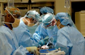
Spine Tumor Surgery in Karachi: Leading the Way with Prof. Dr. Akbar Ali Khan, Best Neurosurgeon in Pakistan

Advancing Spinal Health: Understanding Vertebrectomy with Prof. Dr. Akbar Ali Khan
Spinal Cord Stimulation

Kyphoplasty and Vertebroplasty
Vertebrectomy/Corpectomy

Foraminotomy
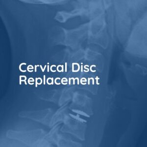
Cervical Disc Replacement

Anterior Cervical Discectomy and Fusion
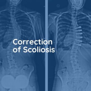
Correction of Scoliosis

Thoracic Discectomy
Lumbar Facet Joint Replacement
Interspinous Spacers

Endoscopic Lumbar Discectomy

Laminectomy
Disc Replacement Operation
Posterior Lumbar Fusion


